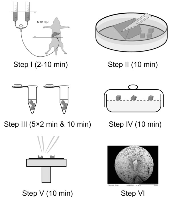
Specimen Preparation For Sem. Some particles have natural tendency to absorb moisture from regul environment. Sample preparations are essential in scanning electron microscopy. Before SEM characterization Clean and dry no outgassing. Ideally the smallest representative sample size is the one to use.

Chromium gold platinum etc for examination under a conventional SEM often referred as ambient temperature scanning electron microscopy ATSEM. For scanning electron microscopy of electrically insulating materials the. Always check recent published research papers to check on current techniques being used. Thus the aim of this chapter is to equip researchers post graduate students and technicians with essential knowledge required to prepare samples for scanning electron microscopy SEM investigations in the life sciences. Mechanical preparation is the most common method for preparing these so-called materialographic or metallographic samples for microscopic examination. For biological samples critical drying to remove liquid without damaging the specimen.
Specimen size depends very much on SEM model you use.
Preparation often involves nothing more than mounting a small piece of the specimen in a suitable liquid on a glass slide and covering it with a glass coverslip. The artifacts may appear as specific features in the backscattered electron mode eg cracks introduced in cutting or drying. Chromium gold platinum etc for examination under a conventional SEM often referred as ambient temperature scanning electron microscopy ATSEM. Proper preparation of flat-polished specimens for SEM is important. This kills the tissue sample at the same time. Standard SEM procedures for biological samples involve chemical fixation dryingdehydration mounting on a stub and coating with a metal eg.